

Key Takeaways
- A visual field test measures your peripheral (side) vision.
- The test is painless and often part of a routine eye exam.
- It can pick up early signs of glaucoma before you notice any change.
- Blind spots, called scotomas, vary in size and shape.
- Repeat testing helps confirm results and track changes over time.
A Simple Test With a Lot to Say About Your Vision
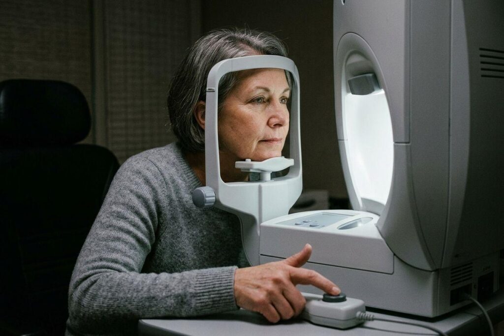
You sit in a dim room, rest your chin on a small cup, and click a button every time a tiny light flickers at the edge of your view. It feels almost like a video game. A few minutes later, your eye doctor at The Eye Gallery has a map of where your eye sees effectively and where it doesn’t.
A visual field test measures your peripheral (side) vision. It’s quick, painless, and gives your optometrist important information. It’s one of several tools used during eye disease diagnosis and management.
What a Visual Field Test Measures
A visual field test measures how well you see things in different areas of your vision. This includes the space above, below, to the left, and to the right of whatever you’re focusing on. Many people go through a visual field test as part of a routine eye exam. Nothing touches your eye during the test, and it should be painless.
Why Your Eye Doctor in Wichita KS May Recommend One
Glaucoma can begin affecting your side vision long before you notice a problem. Your brain simply fills in the gaps. A visual field test can catch these important early changes before glaucoma can progress.
The test also works as a record. By comparing results over months or years, your eye doctor in Wichita KS can see whether your vision is holding steady and whether a treatment plan is doing its job.
Conditions Often Monitored with Visual Field Testing
Visual field testing helps track a handful of concerns. These include:
- Glaucoma and other optic nerve concerns
- Stroke, multiple sclerosis, and thyroid eye disease
- Long-term use of certain medications
Blind Spots & What They Show
Missing areas in your field of vision are called scotomas. Their size and shape matter, because different patterns point toward different underlying concerns. Identifying scotomas can help your eye doctor make a diagnosis.
Types of Visual Field Tests
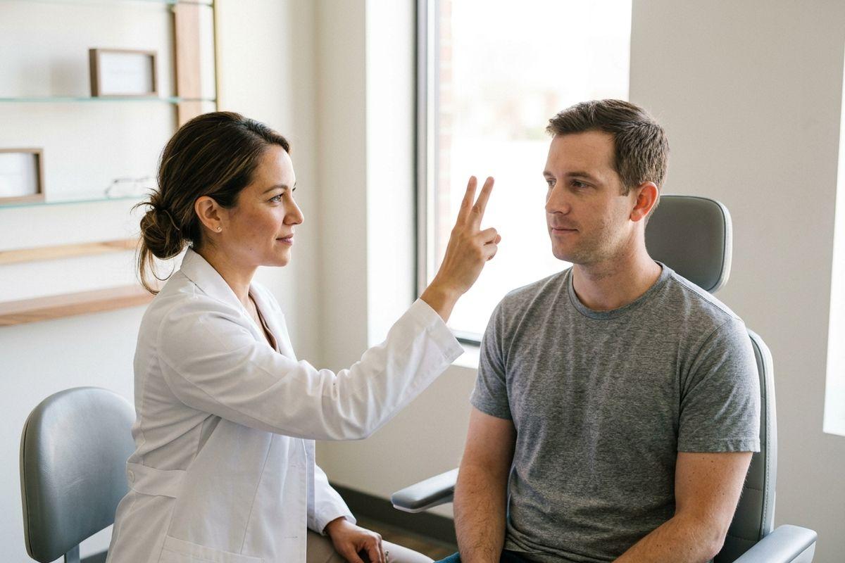
- Confrontation test. Your eye doctor sits in front of you and asks you to count fingers held off to one side. It takes only seconds and gives a quick read of your peripheral vision.
- Automated static perimetry. You look into a machine in a dim room and press a button each time a small light appears. This test produces a detailed map of your peripheral vision that your eye doctor can use to track changes.
- Amsler grid. A small square of lines with a dot in the middle, often used at home to check your central vision between visits. This tool can flag early warning signs of macular degeneration.
What to Expect During the Test
Your job is to keep looking straight ahead at the central target and press a button whenever you notice flashes of light at the edges of your visual field. Blinking is fine, and so is asking for a short break if your eye feels tired.
How to Prepare
Show up rested. Tiredness can drag down your response time and skew the results. Bring your current glasses prescription too, since the right lens power may help make testing easier.
What Happens Afterwards
A single test doesn’t tell the whole story. Repeating the test may be needed to confirm what the first one showed, especially if the results looked unusual.
If the test shows vision loss, this can affect your ability to drive. However, that’s a conversation worth having directly with your eye doctor.
Why the Test Is Worth Your Time
The test finds changes you can’t feel on your own. Paired with optic nerve imaging, it gives your doctor two views of the same picture and a clearer sense of what’s happening over time.
Book Your Next Eye Exam
Ten quiet minutes in a dim room can tell your doctor a great deal about the health of your vision. If it’s been a while since your last checkup, or if glaucoma runs in your family, now is a good time to schedule.
If you’re having vision struggles, our team at The Eye Gallery in Wichita would be glad to see you and your family. Call to book your appointment today.